It is estimated that up to 200,000 people globally die from fungal lung infections (Aspergillosis). Now, the EU-funded Fp7 MATHIAS project has developed an imaging technique to help doctors fight this huge killer of patients with weakened immune systems.
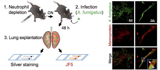
 The new test involves attaching radioactively labelled antibodies to the infecting structures formed by the fungus Aspergillus fumigatus.
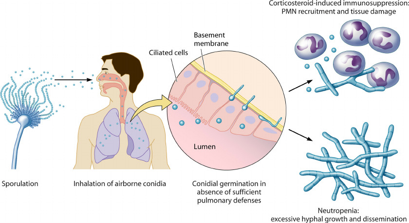
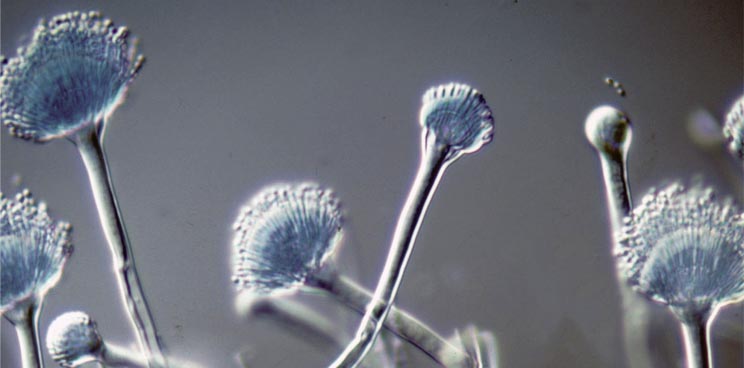
These fungal spores (conidia) are inhaled every day and do not usually cause a problem for healthy individuals. However, in patients who are immunocompromised (e.g. due to leukaemia or bone marrow transplants etc.) they can settle in the lungs and develop into invasive pulmonary aspergillosis.

At present, a definitive diagnosis for the disease is only obtained at autopsy or relies heavily on an invasive biopsy – an extremely unpleasant procedure which is not always applicable in suffering patients.
By using radioactively labelled antibodies in the new imaging method, clinicians researchers were able to image the growing fungus in real-time.
The project researchers used a combination of PET and MRI imaging (diagnostic tools that are available in most hospitals) to identify the disease and to rule out lung infections caused by other pathogens, such as bacteria or viruses.

The project, which will run until 2018, is also working on developing new treatment options which can replace the systemic anti-fungal drugs that are currently administered to patients but are known to provoke severe side effects.
A small-scale clinical trial will then be undertaken in Germany, in full compliance with the Declaration of Helsinki on Good Clinical Practice and relevant EU and German clinical trial regulations.
This infectious disease is one of the common causes of death in immunocompromised patients and exerts a tremendous cost on European healthcare systems. The Mathias project could therefore really radicalise diagnosis of fungal lung infections.


