


Newsletter Signup - Under Article / In Page
"*" indicates required fields
Although we have come very far in understanding cells and their functions, many undiscovered characteristics still keep us in the dark. Creating experiments that mimic the cell’s in vivo environments as closely as possible is one of the many challenges researchers face when working with cells in their labs.
Micropatterning is a technique that addresses this challenge. It combines the traditional cell culture substrates with the physiological relevance of in vivo experiments, building in vitro microenvironments similar to those of the cells’ in vivo situations. The technique allows researchers to control cellular patterns and increases the relevance and repeatability of the data generated, while decreasing the requirement for experiments on animals.
The creation of microenvironments can be used to form an understanding of the natural relationship between tissue structures and functions, and can give clues on how this relationship can become unbalanced in case of disease.
Creating microenvironments helps research

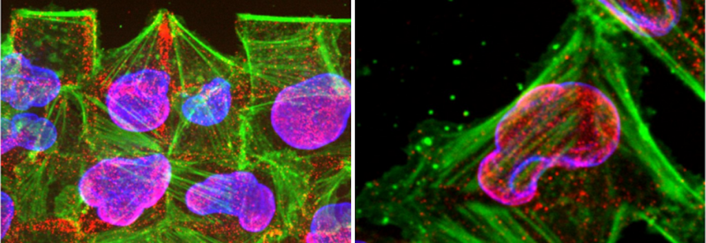
As opposed to traditional growth media, the creation of a microenvironment through micropatterning allows researchers to manipulate their cell cultures on a very refined scale. Different parameters, such as the biochemical and mechanical nature of the substrate, can be tweaked to observe the cell’s reactions to different environmental factors.
Cells are highly sensitive to their mechanical, geometrical and biochemical environments, so these changeable parameters allow the observation of cellular and tissue biology, including cell survival, proliferation, differentiation, migration, and cytokinesis.
Most micropatterning techniques are based on the same three steps. At first, researchers have to generate a pattern with proteins from the extracellular matrix (ECM) on their substrate of choice. Then, the so-called seeding of a cell population occurs, where cells ‘stick’ to the manipulated substrate with a controlled positioning and adhesion. Lastly, a washing process removes excess cells and reveals the pattern of choice.
How does micropatterning work?
Among other micropatterning techniques, additive micro-contact printing is probably the most famous one. Here, stamps with so-called micrometric motifs are prepared with an adhesion biomolecule and then transferred onto the substrate of interest.
Micro-contact printing allows the direct control of cell adhesion, however it comes with several disadvantages: The stamp only allows one type of protein to be transferred at a time and the user cannot control the density of the biomolecule either. Also, the stamp has to be built prior to its use in a complex process, making it time-consuming and quite costly.
Alvéole is a company that has risen to the challenge of creating a micropatterning technique which addresses these disadvantages. It’s main technology PRIMO is designed to meet the need for better controlled cell cultures.
A revolution in cell biology
“Cell biology is undergoing a revolution. It is now well-known that the way cells are grown, namely in plastic, flat, Petri dishes does not recapitulate the conditions in which cells live in vivo,” explains Pierre-Olivier Strale, senior scientist at Alvéole. “Creating more biomimetic environments is of critical importance since the discoveries made from the fundamental research is driving drug development and clinical trials.”
PRIMO is a contactless micropatterning method based on light-induced molecular adsorption of Proteins (LIMAP) technology, which relies on the combined action of UV light and a photosensitive reagent. Together with the UV-light, the photosensitive reagent can activate the substrate specifically where the biomolecule of interest should attach.
Contactless micropatterning with UV light
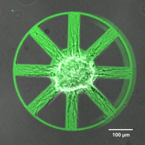
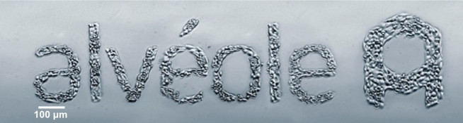
A UV laser beam shines onto a surface made of thousands of micromirrors. Each micromirror is independent and can be controlled by the software that Alvéole has developed, called LEONARDO. This program allows the user to basically upload or draw any image they want, it will then project the shapes of light directly onto the cell culture substrate through the objective of the microscope.
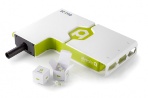
“PRIMO is compatible with the majority of microscopes on the market, so people will already have part of the technology in the lab,” describes Pierre-Olivier Strale. “It’s a bit like a projector which you use to shine a movie onto the wall.”
It is well known that in vivo cells often respond to different molecule concentrations and to the stiffness of their environment. Therefore, Alvéole developed PRIMO which, unlike traditional technologies, allows the creation of protein gradients on all the standard cell culture substrates, whether stiff or soft, flat (2D) or microstructured (3D).
Research applications of PRIMO
Furthermore, PRIMO enables the printing of multiple proteins, rather than only one, allowing the generation of complex co-cultures. It can also control the density of molecules on a substrate, which is useful for cell-migration studies.
PRIMO’s largest advantage over traditional micropatterning technologies is its flexibility. “When using our technology researchers are not forced to build a stamp, but rather they simply load an image into our software,” says Pierre-Olivier Strale. “You can start an experiment in the morning, realize it’s no good and then reload an updated version within minutes. The micromirrors will shape the UV light and locally activate the cell culture surface.”
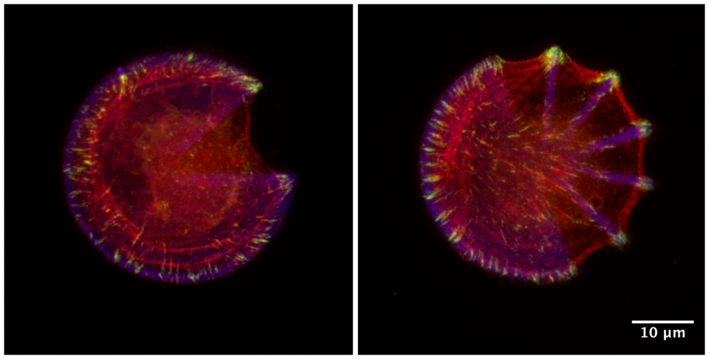
The high resolution achieved by PRIMO – 10 to 20 times smaller than a single cell – allows researchers to control the parameters of the microenvironment in a very defined manner. This is important, as most users are trying to model disease with the goal of understanding the evolution of diseases in the fields of oncology, hepatology and cardiology in order to develop drugs.
Another application of PRIMO is in fundamental research. Researchers are looking at creating artificial tissues for regenerative medicine purposes. Alvéole is currently collaborating with many international researchers, which use Alvéole’s latest innovations, including PRIMO, to test them on novel experiments and then give their feedback on the results.
“I believe that our company can create new research tools for cell biology and accompany the researchers in the long term,” explains Romuald Vally, CEO of Alvéole. “By combining the expertise and the enthusiasm of our team and our high-end technology platform, we are creating new practices in the cell microenvironment market.”
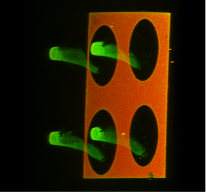
Controlling cells, a new kind of art
One look at the patterns created by Alvéole’s PRIMO makes you think of art. “With PRIMO there is no limitation in terms of the shapes of the molecules that can be attached to the surface, the only limitation is your imagination,” agrees Pierre-Olivier Strale. “It’s like painting with proteins and cells.” He does, however, emphasize that PRIMO is first and foremost a tool for scientific research and the artwork is just “a happy side-effect.”
Want to bring your research further and leave suboptimal micropatterning techniques behind? Get in touch with Alvéole and learn more about their high resolution photopatterning technology PRIMO!
Title Image: Damian Ryszawy/ Shutterstock
Article Images via Alvéole
Video 1: Video (24h time-lapse): Axon guidance on the spokes and around the circumference of the laminin wheel pattern (Source: H. Ducuing, R. Moore, Y. Lecomte, P.-O. Strale, V. Studer)